



Among 260 patients who underwent ultra low dose chest CT screening, 179 subsequently had biopsies based on imaging findings and 54 cancers were identified, while a separate coronary CT programme used approximately 15 mL of contrast per examination.
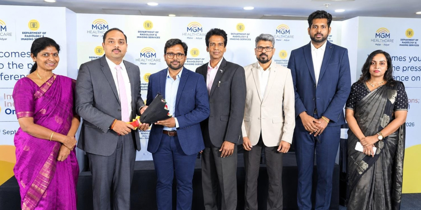
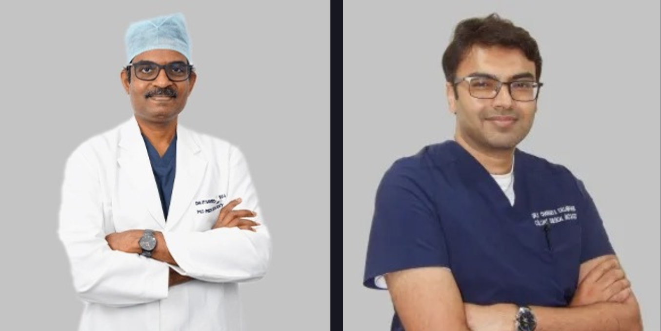
Dr. Samuel Reefath J, Senior Consultant & Lead Radiologist, and Mr. Venugopal Bhat, Chief Operating Officer and Group Vice President, Strategic Initiatives, say MGM Healthcare Malar Adyar is using low dose CT and Deep Learning to reduce patient exposure while supporting earlier and more precise diagnosis
Lower radiation and contrast exposure is becoming part of CT based assessment at MGM Healthcare Malar Adyar, where new imaging protocols are being used for lung cancer screening and coronary artery evaluation.
The hospital has introduced ultra low dose CT chest screening and low dose CT coronary angiography alongside Deep Learning based image reconstruction and quantitative imaging analysis. The approach is intended to retain the diagnostic information required by clinicians while reducing radiation exposure and, in selected applications, the amount of contrast used during examinations.
A central component of the CT workflow is Delta, a Deep Learning based image reconstruction technology. It is being used to reconstruct diagnostically useful images from scans acquired at substantially lower radiation doses.
The hospital said 133 coronary CT examinations were performed over a 10 day period using approximately 15 mL of contrast for each examination and a low radiation acquisition strategy. The approach is intended to reduce radiation and contrast exposure while maintaining the image quality required for assessment of the coronary arteries.
MGM Healthcare Malar Adyar has also used its ultra low dose chest CT programme for 260 patients. Based on imaging findings, 179 of these patients subsequently underwent biopsy, with 54 cancers identified within the screened group.
Dr. Samuel Reefath J, Senior Consultant & Lead Radiologist, said the focus is on obtaining clinically useful information while limiting the burden associated with an examination.
“Every time we perform a CT examination, we have to ask two questions: Can we obtain the information the clinician needs? And can we do it with the least possible burden to the patient? That burden includes radiation, iodinated contrast, cost, time and sometimes even the anxiety associated with invasive investigations,” Dr. Reefath said.
“We therefore wanted to move from a conventional approach of simply acquiring images towards precision imaging, obtaining the maximum clinically useful information with the minimum necessary exposure. This philosophy has driven our low-dose coronary CT, ultra-low-dose chest CT and our work in quantitative and AI-assisted imaging. We are not abandoning established CT principles. We work within accepted radiology and cardiovascular CT standards, while optimizing acquisition parameters for individual clinical situations.”
Deep Learning is also being incorporated into the analysis of imaging data to assist radiologists with identifying findings that may require closer assessment, organising information and quantifying abnormalities.
“Deep Learning-based tools can support the identification of subtle abnormalities, assist in image analysis, and help organise and quantify information, allowing the radiologist to focus more closely on the clinical interpretation of the findings,” Dr. Reefath added.
Mr. Venugopal Bhat, Chief Operating Officer and Group Vice President, Strategic Initiatives, said the hospital is focusing on how imaging technology can contribute to earlier and safer diagnosis.
Our vision is to move towards precision healthcare rather than simply more healthcare technology. Advanced imaging should ultimately answer three questions: Can we detect disease earlier? Can we characterise it more accurately? And can we do it more safely?” Mr. Bhat said.
He added that the value of imaging technology ultimately depends on its benefit to patients.
“If we can detect cancer before symptoms develop, that potentially creates an opportunity for earlier treatment. If we can perform a coronary CT with substantially less contrast and radiation, we reduce the burden of the investigation. And if quantitative imaging and AI can help us extract information that is difficult to appreciate visually, we can potentially provide clinicians with more objective information. So, the final measure of innovation is not how sophisticated the scanner is. The final measure is whether the patient benefits,” Mr. Bhat said.
At Prittle Prattle News, featuring you virtuously, we celebrate the commitment and innovation. Led by Editor-in-Chief Smruti Bhalerao, our platform is dedicated to sharing impactful stories that inspire change and create awareness. Follow us on LinkedIn, Instagram, and YouTube for more stories that matter.
About Prittle Prattle News
Prittle Prattle News is a digital news publication covering business, technology, healthcare, education, lifestyle, public affairs and other developments from India and around the world. Led by Editor-in-Chief Smruti Bhalerao, we publish timely news, industry updates and feature stories for a broad readership.







Add Comment